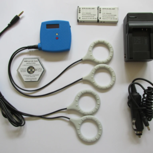
StemPulse and MultiPulse – Great science leads to great health!
The story of StemPulse and MultiPulse is the story of David and Goliath. Dr. Bob Dennis, Ph.D., Biomedical Engineering, invented the ICES technology that powers StemPulse and MultiPulse. He was just an engineer; working for NASA, MIT and the University of N. Carolina when he was introduced to PEMF. Dennis built PEMF machines to experiment with stem cells by using multiple frequency patterns to excite cells and make them stronger.
Dr. Dennis really didn’t understand the widespread use of PEMF…
…until he used it on himself!
The result was a transformation.
Suddenly, he wanted to make PEMF machines that anyone could afford. Perhaps, Dennis’ friends told him “This is a big industry with lots of established brands. You can’t beat them!” Yet, Dennis’ went forward just like David at the Valley of Elah. Arguably, going from engineer to product developer and manufacturer isn’t the same as slaying giants. However, Dr. Dennis has used small size and sharp tactics to take on bigger and way more expensive machines. Furthermore, he has become very successful and continues to develop highly advanced PEMF devices such as MultiPulse.
Watch the video to hear the story in his own words:
Bob Dennis PhD talks about why he developed miniature PEMF
View the MultiPulse in the video here
View Product
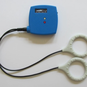
StemPulse was the first device designed as the miniature giant slayer. Conversely, it delivers a strong and reliable magnetic signal at up to 20,000 microtesla (uT). Importantly this is done using the 2 coil system to amplify the healing power of PEMF . Simply, the StemPulse uses efficiency to create the same treatment conditions that larger and much more expensive machines offer.

20,000 microtesla (200 Gauss) using this array
StemPulse delivers at 1/50th the size and 1/10th the cost!
Many scientific PEMF claims are based on one study on a few patients. On the other hand, Dr. Dennis has proven many times over that his machines invigorate cell activity that promotes healing. StemPulse and MultiPulse are backed by repeatable scientific experiments. Additionally, independent scientists found increased stem cell growth and double the production of extracellular matrix (ECM); the connective structure between cells.
Most importantly, this stimulation would allow rapid ‘self repair’ throughout your whole body. In other words, using it on your knee or hip, positively affects other areas of your body. With this in mind, you can see that it is much like when David took on Goliath. A very small action may cause a very large result.
Compare StemPulse and MultiPulse
The two devices are similar in many ways. Both are quite small and reasonably priced. Additionally, they both use the same applicators. Conversely, there are distinct differences.
Basically, MultiPulse can deliver almost every treatment protocol that Dr. Dennis has considered. Brain waves, Schumann resonance, simple pulses and custom frequencies developed for experimentation. All of these have been tested in the lab or clinically and many are the same as offered by larger, more expensive machines. Also, MultiPulse has special protocols available only on these machines. These are effects which address the 3-dimensional nature of cells.
The StemPulse only has only one protocol, optimized for severe chronic pain and orthopedic injury. However, it has also been tested successfully on traumatic brain injury and renal insufficiency in cats. Importantly, this indicates the variable signal produced is effective beyond the expectations of it’s users. Likewise, we continue to see these effects in all PEMF use cases;i.e., performance better than expected.
StemPulse has other advantages such as an easy to find power source (9v battery) and it can run two applicators at a time. Otherwise, you may opt for the MultiPulse’s many specialized applications such as sleep or recovery. Also, the MultiPulse is the smallest with the longest battery life. This makes it great for work or travel. Either way, you will find a variety of uses and excellent performance from both devices.
Technical Information
Features
| StemPulse | MultiPulse |
| Pulse Generator StemPulse | Pulse Generator MultiPulse |
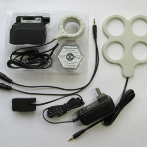
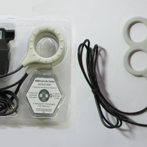
| One pair of Short Coils 12” | One pair of Short Coils 12” |
| One Pair of Standard Coils 20″ | One pair of Standard Coils 20″ |
| One 9V battery (not rechargeable) | One Coil Life Test Chip |
| One Coil Life Test Chip | Two DLI88 compatible camcorder batteries (gift) not included for international orders due to FAA regulations |
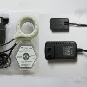
| 12 months manufacturer warranty for Pulse Generator (Warranty doesn’t cover coils, water damage or physical damage) | One DLI88 battery charger with car adapter (gift) not included for international orders due to FAA regulations |
| 30-day return period ($100 restocking fee) | 12 months manufacturer warranty for Pulse Generator (Warranty doesn’t cover coils, water damage or physical damage) |
| 30-day return period ($200 restocking fee) |
Performance
| StemPulse | MultiPulse |
| The StemPulse only has only one protocol, the “Omni 8”, which is a sequence of pulses of defined shape with pulse frequencies each applied for several minutes in the following order 5 pulses per second (pps), followed by 100 (pps), 3.9 (pps), 7.1 (pps), 10.4 (pps), 13.7 (pps), and 16.9 (pps). When complete, this cycle of pulses repeats continuously. | Standard and Legacy ICES Protocols — B5 – C5, — A9, — P2 (SomaPulse, AllevaWave, …),– Omni 8 Schumann resonances and harmonics (pulses per second) — Schumann 1 –(7.83 pps), — Schumann 2 –(7.83, 14.3 pps),– Schumann 3 –(7.83, 14.3, 20.8 pps), — Schumann 4 –(7.83, 14.3, 20.8, 27.3 pps), — Schumann 5 –(7.83, 14.3, 20.8, 27.3, 33.8 pps) Fixed (constant) pulse rate protocols — 1 pps –continuous, — 2 pps –continuous, — 3 pps –continuous, — 4 pps –continuous, — 5 pps –continuous, — 10 pps –continuous TMS protocols (Transcranial Magnetic Stimulation) — scTMS 10pps 30 minutes, — scTMS 10pps 60 minutes Brainwave Entrainment Protocols –alpha wave, –beta1 wave –(low range), –beta2 wave (mid range), –beta3 wave (high range), –delta wave, –theta wave, –mu wave, –SMA wave, –gamma wave Standard Protocols with 5 minute rest between cycles: — B5 –C5 –REST 5, — A9 –REST 5, — P2 –REST 5, — Omni 8 –REST 5 |
Coil Placement
 |
 |
Using the MultiPulse (Video)
Prices and Packages
| StemPulse | MultiPulse |
| StemPulse with all standard features 429.00 | MultiPulse with Standard Features 629.00 |
| StemPulse with all standard features and 9V Adapter 464.00 | MultiPulse with Standard Features and 120AC Adapter 647.00 |
| StemPulse with all standard features and Box Coil Array 499.00 | MultiPulse with Standard Features and Box Coils 699.00 |
| StemPulse Complete Set with Adapter and Box coils 534.00 | MultiPulse System Complete Set with adapter and box coils 717.00 |
StemPulse and MultiPulse Accessories
Here Is How I Use The StemPulse & MultiPulse For Work And Great Sleep!
Life Saving Power for Pets
PEMF manufacturers guard against attacks by the FDA and others by not mentioning anecdotal data. Strictly, people just using PEMF and seeing great results is not enough to prove the efficacy of medical devices. However, our StemPulse makers don’t mind including animals in their story. Fortunately, we found a study on feline kidney disease and used it successfully on the office cat.
From Russ Guillemot (the boss):

“Casey, was born in my closet in 2003 (she’s 18). She is an indoor, upstairs cat. She was sick every day; making a mess of the carpet and barely eating.
Sadly, according to the Journal of Feline Medicine and Surgery, renal failure is a leading cause of suffering and death in domestic cats, with approximately 1 in 3 cats affected. I was really worried.
Great news came from an internet search. Surprisingly, the StemPulse was used successfully to treat a cat with the same symptoms. The same machine I use almost every day! The report chronicles the return to normal and then reversion to renal insufficiency in a single cat, when PEMF was applied or withheld. This happened over three cycles of application and non-application over a 5-year period.
Oct 3, 2020 I put a PEMF coil on Casey and under her bed 24/7. In the next 30 days she was sick only 3 times. 3 months later she has no symptoms as if she is completely cured.
Happy cat! Happy owner!!” Read the full story, videos and more pictures of Casey here.
Bone Growth Stimulator
Rabbit osteotomy study. This is work conducted at Texas A&M on New Zealand White Rabbits using the StemPulse signal.

A number of rabbits had incisions made in the forearm bone, the ulna, where section of the bone was removed leaving a significant gap. Under normal circumstances this gap will not heal in, because of the extent of the gap. In the control group without PEMF stimulation, what would normally happen was revealed, that is, there was no bone regeneration in the gap. In the treatment group, after 14 days of treatment there was clear evidence of bone growth in the gap. After 28 days, in the untreated group, there was still no bone growth in the gap. However, in the StemPulse treated group, there was a dramatic increase in bone growth, and tendons and ligaments.
This technology clearly increases healing in muscle, tendon, ligament, skin, and bone tissues. The study was terminated because the researchers could clearly see the difference between the active PEMF system and the sham control devices.
Click the link below to see all the images of the rabbit’s leg bone regrow.
Rabbit Regrowth Progression

Osteotomy incision in rabbit..

Xray of osteotomy incision in rabbit.

Osteotomy incision in rabbit 14 days after without PEMF. No change.

Osteotomy incision in rabbit 28 days after without PEMF. No change.

Osteotomy incision in rabbit 14 days after with PEMF. Huge difference.

Osteotomy incision in rabbit 28 days after with PEMF. Bone fully regrown.
The Science Behind, and History of the Stempulse
The Patented Science of This Technology and History of the Stempulse
The Patented Science of This Technology and History of the Stempulse
History of the Stempulse
This new American-developed technology is the result of scientific research and development that evolved from programs funded by NASA (National Aeronautics and Space Administration) in the 1990?s. Dr. Robert Dennis was the design engineering and biophysics consultant to the NASA program and has continued to develop the technology under an exclusive sub-license of the NASA patents. After more than 15 years of research and development of the PEMF technology, radical design enhancements, and several new patents beyond the original NASA research, this technology is now being introduced for use with both humans and animals.
This technology has been shown to create nerve stem cell fiber growth in culture and to double the rate of production of ECM (extracellular matrix – the connective structure between cells) in at least two scientific studies. Testing has shown significant reduction in joint pain within a few days to weeks when used as directed.
Principles of Operation
The design of the StemPulse device is based on known laws of classical physics from Maxwell’s equations, specifically Faraday’s Law of Induction, as well as both classical quantitative physiology of excitable tissues and modern theories on functional adaptation in musculoskeletal, cardiovascular, and other structural tissues.
It is known that muscle and nerve tissues respond directly to electrical excitation by the trans-membrane cascade of ions; this results in the propagation of electrical action in nerves or in contraction in muscles. It is also widely thought that most structural tissues, especially bone, tendon, ligament, articular cartilage, and perhaps skin, respond to selective ion flow in the paracellular space between the cell membrane and the extracellular matrix (ECM) when the tissue is subjected to mechanical stress. This ion flow is thought to be the signal that initiates the cellular-level response leading to functional adaptation, repair, and regeneration of structural tissues.
The StemPulse device works by using Faraday’s law of induction which says that the leading-edge of the magnetic field is proportional to the induced electric field. That means that the front part of the magnetic field causes an increase in the energy in the cell, i.e. the charge. It is this law that makes things work like electrical transformers, electric motors, and magnetic audio and video tape. Based on these known laws, StemPulse, has the type of magnetic field necessary to generate an electrical field in the para-cellular space around each cell in the right range of field strength and duration to cause a cellular response. These calculations are based on classical physiology where amplitude is equal to Rheobase (intensity) and duration is equal to Chronaxie (time).
There are at least 800 peer-reviewed papers, supporting the fact that electrical shock and electrical currents can induce significant acceleration of restoration in bone tissue and other tissues (tendon and ligament). But the problem has always been (a) locating electrodes close enough to and properly placed around the bones to be stimulated electrically, and (b) the fact that free electrical current follows a random path of least resistance resembling a lightning bolt. A few cells directly in the path of the “lightning bolt” will be hyper-stimulated, while the other 99% of cells outside the “lightning bolt” receive essentially no stimulation. Yet crude as it is, electrical stimulation of bone still has significant beneficial effects when applied properly. The scientific literature in support of this conclusion is very compelling.
To solve these two problems the StemPulse device uses the application of magnetic fields as opposed to electrical fields. It applies magnetic fields (not direct electrical stimulation) mainly because PEMFs do NOT interact directly with the tissues of the body. And the device rapidly changes the strength of the magnetic field. The magnetic field is applied to a volume of tissue using two small coils and then rapidly changing the magnetic field so that it has the optimum level of induced electrical field amplitude (“Rheobase”, calculated from Faraday’s equation) and optimum duration (“Chronaxie”).
Fundamentals of the StemPulse PEMF Technology
Uniform cell stimulation
No electrodes, non-invasive
Tissue subjected to minimum stress
Safe, low-power system
Pulse output tuned to be truly effective
Signal based on extensive science
Based on NASA and additional patents
… see more under Stempulse tab
Patented technology to accelerate tissue healing
The StemPulse technology, originally invented at NASA, employs a carefully designed sequence of magnetic pulses programmed to provide a Pulsed ElectroMagnetic Field (PEMF) around a volume of tissue that is experiencing pain or to assist in the healing process. Currently this device is designed to safely promote healing in muscle, tendon, ligament, skin, and bone tissues. These tissues make up more than 75% of the weight of the body of animals and humans, and often in one or more of these tissues debilitating injury and pain need to be corrected. The StemPulse is unique because it is based on the physics of electricity and magnetism as well as the physiology of cells and tissues.
It is fair to say that most of the available competing technologies are based on a “informed guesses” and design ideas. Engineers are then hired to produce the theoretical or envisioned signal. But that is simply not good enough. The approach used with the StemPulse has been to use physics and physiology as a guide to the design of the device, and to employ new scientific experiments as needed to fill in the gaps of knowledge, allowing the StemPulse to become commercially available as a very effective and lower cost and portable, battery-operated. Continued focus on the science and engineering and strategic partnering for product testing and development with selected members of our customer base have led to this current version of the StemPulse P1 device, both for human and veterinary use.
THE SCIENCE TO DATE
Below is a summary of the research that initiated the development of the StemPulse device, starting with the stimulation of neural stem cells for NASA. Dr. Robert Dennis, PhD, chairman of the department of biomedical engineering at the University of North Carolina, was the inventor of the device used in this study.
Physiological and molecular genetic effects of time varying electromagnetic fields on human neuronal cells
Thomas J. Goodwin, PH.D., Lyndon B. Johnson Space Center
National Aeronautics and Space Administration (NASA), Johnson Space Center, Houston, Texas
The present investigation details the development of model systems for growing two- and three dimensional human neural progenitor (NHNP ) stem cells within a culture medium facilitated by a time-varying electromagnetic field (TVEMF), ie PEMF. The cells and culture medium are contained within a two- or three-dimensional culture vessel, and the electromagnetic field is emitted from an electrode or coil. These studies further provide methods to promote neural tissue regeneration by means of culturing the neural cells in either configuration. Grown in two dimensions, neuronal cells extended longitudinally, forming tissue strands extending axially along and within electrodes comprising electrically conductive channels or guides through which a time-varying electrical current was conducted. In the three-dimensional aspect, exposure to TVEMF resulted in the development of three-dimensional aggregates, which emulated organized neural tissues. In both experimental configurations, the proliferation rate of the TVEMF cells was 2.5 to 4.0 times the rate of the non-waveform cells. Each of the experimental setups resulted in similar molecular genetic changes regarding the growth potential of the tissues as measured by gene chip analyses, which measured more than 10,000 human genes simultaneously. This study clearly the ability to use TVEMF to control the proliferative rate, directional attitude, and molecular genetic expression of normal human neural progenitor cells. The procedure is applicable to, but not limited to, the control of NHNP cells in both two-dimensional and three-dimensional culture. The genetic responses both up-regulated and down-regulated genes which were maturation and growth regulatory in nature. These genes are also primarily involved in the embryogenic process. Therefore it is reasonable to conclude that control over the embryogenic development process may be achieved via the presently demonstrated methodology. Specific genes such as human germline oligomeric matrix protein, prostaglandin endoperoxide synthase 2, early growth response protein 1, and insulin-like growth factor binding protein 3 precursor are highly up-regulated. Keratin Type II cytoskelatal 7, mytotic kinesin like protein 1, transcription factor 6 like 1, mytotic feedback 27 control protein, and cellular retinoic acid binding protein are down-regulated. Each of these two sets is only an example from the approximately 320 genes changes expressed as a consequence of exposure to TVEMF.
There is significant precedent in the literature for the results reported above. Kepler et al. (1990) reported the effects of the neurons with oscillatory properties on the composite of neural networks. This work illustrates the likelihood that a pulse width modulated system might bring on specific responses in neural tissues. As previously discussed, Valentini et al. (1993) demonstrated the ability to enhance the outgrowth of neural fibers on materials that possess a weak electric charge. This would indicate that intense electric fields are not necessarily an essential component of this process, and that a weak and persistent stimulus might yield a measurable effect.
Additional evidence of the effects of magnetic fields exists in the work of Sandyk et al. (1992a). This communication details dramatic improvement of a patient with progressive degenerative multiple sclerosis. Briefly, the patient showed considerable improvement when subjected to treatment at a frequency of 2-7 Hz and an intensity of the magnetic field of 7.5 pico Tesla. These parameters marginally parallel those of this report. In a similar fashion, Sandyk et al. (1992b) reported significant improvement in patients treated with the same field strength and intensity. The ability to suppress or stimulate the growth of non-excitable cells has been reported in mouse lymphoma cells by Lyte et al. (1991). A narrow range of electric field was found to be effective at one end to stimulate and at the other to inhibit the growth of these cells. These data might suggest cellular receptors in all cells. To sustain this notion, Brüstle et al. (1996) reported the potential to use neural progenitors for recapitulation of neural tissues. As would be expected, this would require genetic control at the embryonic level. We believe this study indicates our ability to trigger these parametric events.
As is clearly demonstrated in the human body, the bioelectric, biochemical process of electrical nerve stimulation is a documented reality. The present investigation demonstrates that a similar phenomenon can be potentiated in a synthetic atmosphere, i.e., two-dimensionally or in rotating wall cell culture vessels.
One may use this electrical potentiation for a number of purposes, including developing tissues for transplantation, repairing traumatized tissues, and moderating some neurodegenerative diseases and perhaps controlling the degeneration of tissue as might be effected in a bioelectric stasis field.
— From PHYSIOLOGICAL AND MOLECULAR GENETIC EFFECTS OF TIME-VARYING ELECTROMAGNETIC FIELDS ON HUMAN NEURONAL CELLS
Thomas J. Goodwin, PH.D., Lyndon B. Johnson Space Center National Aeronautics and Space Administration, Johnson Space Center, Houston, Texas
.
StemPulse Signal pattern development history
The three stimulation patterns in the StemPulse are based on the stimulation patterns developed over the years for several different applications. Some of this information has been published by the engineer developing the system, some by others, and quite a lot has not been published at all yet. These different types of experiments and applications have driven the understanding of what the best stimulation patterns are likely to be, mainly for skeletal muscle, with the working hypothesis that what is good for skeletal muscle is likely also good for other tissues of the musculoskeletal and neuromotor system, and based on clinical observations with this PEMF system, that seems to be the case.
The different types of experiments and applications worked on with these signal patterns since 1987 include:
- Fiber-type transformation by implantable stimulation for cardiomyoplasty experiments
- Maintenance of skeletal muscle mass and contractility following chronic denervation
- Maintenance of muscle mass and contractility during disuse (hind limb unweighting) experiments
- Excitability and contractility in developing and aging muscle
- Interaction of muscle with tendon and bone tissue during embryonic development (chicks)
- In vitro tissue engineering of skeletal, cardiac, and smooth muscle
- Development of control and maintenance stimulation devices for hybrid prosthetic and robotic devices (having built the first ever hybrid robot powered by living muscle at MIT in 2000-2001)
- Design of in vitro and in vivo bioreactors for growing engineered musculoskeletal and neuromotor tissue cells.
So, taking his experience from about 25 years with all of the above, the most effective and very efficient stimulation patterns were boiled down to three sequences. These electrical stimulation patterns were then transformed by using calculus to determine the necessary magnetic field patterns and how the magnetic fields needed to change over time to induce the desired electrical charge within the tissue being stimulated. Then these magnetic field requirements were converted into a sequence of electrical pulses to be generated by the PEMF generator unit to drive the coils to generate the required magnetic field waveforms. So, it can be seen, that you have to go back several layers to get to the actual stimulation patterns as they would be seen within the PEMF pulse generator itself. These are transformed in space and time to produce the desired fields within the tissue itself. It was also discovered that while a square wave was optimal it was not necessary the most efficient. From an engineering perspective it’s almost impossible to generate a true square wave. Therefore, using engineering assessments, an optimized trapezoidal wave was the results.
From research and experience, it is known that stimulation of muscle will stimulate nerves which stimulates bones and therefore all the intermediately involved cells as well. There are fast twitch and slow twitch muscles. Stimulation patterns for both of these types of muscles are necessary to appropriately stimulate all the muscles of the body, or over those areas where the magnetic field is placed.
In addition, the StemPulse system uses another concept common in magnetic field research in biology, a specific design of the coils. That is, the use of Helmholtz coils. Two circular coils are the basis for delivering the StemPulse signal. A Helmholtz pair consists of two identical circular magnetic coils that are placed symmetrically one on each side of the experimental area along a common axis. Each coil carries an equal electrical current flowing in the same direction. The Helmholtz coil configuration makes the magnetic field field more uniform between the 2 coils. This tends to produce more stable and repeatable results and allows for better results when the fields between the 2 coils are oriented in different dimensions.
Basic research underpinning the development of the StemPulse device.
This research summary is of necessity somewhat complex, because of the complexity of the science. It is likely to be mostly of interest to those who have a scientific or engineering background. Nonetheless, this summary serves to highlight the impressive an in-depth scientific and engineering background that served as the basis for the development of the StemPulse device. This rich and in-depth research background, is not typical of most PEMF devices. Because of that it is reasonable to say that the research background behind the StemPulse far surpasses most other PEMF devices that are commercially available for home use.
Based on the research conducted for NASA, and subsequently for Regenetech, and Dr. Dennis’s extensive research history in cell biology and muscle physiology, several patents were applied for and granted.
United States patent. Dennis et al. Patent number US 8, 029, 432 B2 October 4, 2011
Magnetic system for the treatment of cellular dysfunction of a tissue or an extracellular matrix disruption of tissue.
This patent is for a system, kit, and pet bed for treating tissue using magnetic coils connected to a pulse generator. The system can include a power supply, a bidirectional communication and power port, a microcontroller, a processor, a data storage, and a Helmholtz – like magnetic coil pair. The kit can include a saddlebag and a support strap the pet bed can include bedding, a fabric housing, a Halbach array, and an on/off switch
This patent was implemented because there was a need for an inexpensive portable system for therapeutically treating tissue disease of humans and animals that was noninvasive, easy to apply, and provide quick relief.
The patent provides specifications for the current design of the StemPulse device.
United States patent, Dennis et al. patent number 8, 137, 258 B1. March 20, 2012
Magnetic method for treatment of human tissue
This patent is for a method for treating a human having a condition which can include disposing a 1st electromagnetic coil opposite a 2nd Elektra magnetic coil, forming a very low power electromagnetic coil pair and energizing the electromagnetic coils to produce electromagnetic pulses. A plurality of pulse blasts are generated using a connected pulse generator with a power supply. The plurality of pulse blasts use a variety of wave shapes to therapeutically treat tissue of the human.
This patent was implemented because there was a need for an inexpensive portable system for therapeutically treating tissue disease of humans and animals that was noninvasive, easy to apply, and provide quick relief. The patent describes the use of the device for various health conditions.
The patent provides specifications for the current design of the StemPulse device.
United States patent, Dennis et al. Patent number 8, 137, 259 B1. March 20, 2012
Magnetic method for the treatment of an animal
This patent is very similar to the 2 patents above, with the exception of specificity for animals.
Dr. Dennis’s research
Dr. Dennis’s research spans close to 20 years. His primary body of work has been involved in tissue engineering and regeneration as well as stimulation of tissues for regeneration, repair and recovery. It is this research that led him to develop a less invasive signal, that is, a pulsed electro-magnetic field (PEMF) signal. In this research he discovered that a trapezoidal wave is the optimal way for producing this type of tissue stimulation. The original intent was to develop a square wave signal, but from an engineering perspective this is almost impossible. All square wave signals essentially are trapezoidal to a great degree. Dr. Dennis’s broad research background also helped him to determine the best frequencies to be used, not only for the NASA study and device development, but also since that time and enhancing the stimulation protocols in the StemPulse PEMF system.
Below is a summary of research that he either authored or co-authored.
Generally, his research is in the area of tissue engineering. He also has a significant interest in robotics, modern manufacturing technologies, prosthetics, and bio-hybrid systems. This summary is somewhat technical but is typical for the complexities of bioengineering research.
Skeletal Muscle (myooids, U.S. Patent No. 6,207,451) This was the first tissue system developed in my laboratory at the University of Michigan, and is the basis for our further work in the area of engineered, self-organizing tissue systems in vitro. These skeletal muscle constructs are electrically excitable and generate measurable contractile force when stimulated. We have made excellent progress on several of these critical tissue interface technologies: nerve-muscle co-cultures and muscle-tendon junctions. We were, for example, the first to demonstrate the development of a functional nerve-muscle junction in culture, capable of sustaining the transmission of peripheral axonal action potentials to elicit active synchronized contractions in an engineered muscle. The resulting manuscript is in the final stages of review before submission at the time of this writing.
Bioreactors My most significant supporting technology development is a bioreactor system (U.S. Patent Nos. 6,114,164 and 6,303,286). I have also developed and published the designs for electrical stimulator units, specifically designed for use with musculoskeletal tissue, and optical force transducers and other sensors for use in the bioreactor systems.
The potential applications of this research are very far reaching, and include: in vitro tissue models for research in developmental biology aging, injury, and gene therapy; engineered tissue for surgical repair; hybrid robotics, prosthetics, and biomechatronic devices.
Articles with Dr. Dennis as the primary or senior author/investigator
Dennis RG, Whitmire RN, Jackson RT. Action of inflammatory mediators on middle ear mucosa. A method for measuring permeability and swelling. Arch Otolaryngol. 1976 Jul;102(7):420-4. PubMed PMID: 938323.
Miller SW, Dennis RG. A parametric model of muscle moment arm as a function of joint angle: application to the dorsiflexor muscle group in mice. J Biomech. 1996 Dec;29(12):1621-4. PubMed PMID: 8945661.
Dennis RG. Bipolar implantable stimulator for long-term denervated-muscle experiments. Med Biol Eng Comput. 1998 Mar;36(2):225-8. PubMed PMID: 9684464.
Dennis RG, Kosnik PE II. Excitability and isometric contractile properties of mammalian skeletal muscle constructs engineered in vitro. In Vitro Cell Dev Biol Anim. 2000 May;36(5):327-35. PubMed PMID: 10937836.
Dennis RG, Kosnik PE II, Gilbert ME, Faulkner JA. Excitability and contractility of skeletal muscle engineered from primary cultures and cell lines. Am J Physiol Cell Physiol. 2001 Feb;280(2):C288-95. PubMed PMID: 11208523.
Kosnik PE, Faulkner JA, Dennis RG. Functional development of engineered skeletal muscle from adult and neonatal rats. Tissue Eng. 2001 Oct;7(5):573-84. PubMed PMID: 11694191.
Dennis RG, Dow DE, Faulkner JA. An implantable device for stimulation of denervated muscles in rats. Med Eng Phys. 2003 Apr;25(3):239-53. PubMed PMID: 12589722.
Borschel GH, Kia KF, Kuzon WM Jr, Dennis RG. Mechanical properties of acellular peripheral nerve. J Surg Res. 2003 Oct;114(2):133-9. PubMed PMID: 14559438.
Dow DE, Cederna PS, Hassett CA, Kostrominova TY, Faulkner JA, Dennis RG. Number of contractions to maintain mass and force of a denervated rat muscle. Muscle Nerve. 2004 Jul;30(1):77-86. PubMed PMID: 15221882.
Herr H, Dennis RG. A swimming robot actuated by living muscle tissue. J Neuroeng Rehabil. 2004 Oct 28;1(1):6. PubMed PMID: 15679914
Baar K, Birla R, Boluyt MO, Borschel GH, Arruda EM, Dennis RG. Self-organization of rat cardiac cells into contractile 3-D cardiac tissue. FASEB J. 2005 Feb;19(2):275-7. Epub 2004 Dec 1. PubMed PMID: 15574489.
Dow DE, Faulkner JA, Dennis RG. Distribution of rest periods between electrically generated contractions in denervated muscles of rats. Artif Organs. 2005 Jun;29(6):432-5. PubMed PMID: 15926976.
Birla RK, Borschel GH, Dennis RG. In vivo conditioning of tissue-engineered heart muscle improves contractile performance. Artif Organs. 2005 Nov;29(11):866-75. PubMed PMID: 16266299.
Birla RK, Huang YC, Dennis RG. Development of a novel bioreactor for the mechanical loading of tissue-engineered heart muscle. Tissue Eng. 2007 Sep;13(9):2239-48. PubMed PMID: 17590151.
Dennis RG, Dow DE. Excitability of skeletal muscle during development, denervation, and tissue culture. Tissue Eng. 2007 Oct;13(10):2395-404. PubMed PMID: 17867927.
Birla RK, Huang YC, Dennis RG. Effect of streptomycin on the active force of bioengineered heart muscle in response to controlled stretch. In Vitro Cell Dev Biol Anim. 2008 Jul-Aug;44(7):253-60. Epub 2008 Jun 21. PubMed PMID: 18568374.
Dennis RG, Smith B, Philp A, Donnelly K, Baar K. Bioreactors for guiding muscle tissue growth and development. Adv Biochem Eng Biotechnol. 2009;112:39-79.PubMed PMID: 19290497.
Articles with Dr. Dennis as a contributor
Hinds S, Bian W, Dennis RG, Bursac N. The role of extracellular matrix composition in structure and function of bioengineered skeletal muscle. Biomaterials. 2011 May;32(14):3575-83. Epub 2011 Feb 13. PubMed PMID: 21324402;
Donnelly K, Khodabukus A, Philp A, Deldicque L, Dennis RG, Baar K. A novel bioreactor for stimulating skeletal muscle in vitro. Tissue Eng Part C Methods.2010 Aug;16(4):711-8. PubMed PMID: 19807268.
Stemra S, Gilmont RR, Dennis RG, Bitar KN. Bioengineered internal anal sphincter derived from isolated human internal anal sphincter smooth muscle cells. Gastroenterology. 2009 Jul;137(1):53-61. Epub 2009 Mar 27. PubMed PMID:19328796.
Dow DE, Cederna PS, Hassett CA, Dennis RG, Faulkner JA. Electrical stimulation prior to delayed reinnervation does not enhance recovery in muscles of rats. Restor Neurol Neurosci. 2007;25(5-6):601-10. PubMed PMID: 18334775.
DuBay DA, Choi W, Urbanchek MG, Wang X, Adamson B, Dennis RG, Kuzon WM Jr, Franz MG. Incisional herniation induces decreased abdominal wall compliance via oblique muscle atrophy and fibrosis. Ann Surg. 2007 Jan;245(1):140-6. PubMed PMID: 17197977
DuBay DA, Wang X, Adamson B, Kuzon WM Jr, Dennis RG, Franz MG. Mesh incisional herniorrhaphy increases abdominal wall elastic properties: a mechanism for decreased hernia recurrences in comparison with suture repair. Surgery. 2006 Jul;140(1):14-24. PubMed PMID: 16857438.
Borschel GH, Dow DE, Dennis RG, Brown DL. Tissue-engineered axially vascularized contractile skeletal muscle. Plast Reconstr Surg. 2006 Jun;117(7):2235-42. PubMed PMID: 16772923.
Larkin LM, Van der Meulen JH, Dennis RG, Kennedy JB. Functional evaluation of nerve-skeletal muscle constructs engineered in vitro. In Vitro Cell Dev Biol Anim. 2006 Mar-Apr;42(3-4):75-82. PubMed PMID: 16759152.
Arruda EM, Calve S, Dennis RG, Mundy K, Baar K. Regional variation of tibialis anterior tendon mechanics is lost following denervation. J Appl Physiol. 2006 Oct;101(4):1113-7. Epub 2006 May 25. PubMed PMID: 16728516.
Dow DE, Carlson BM, Hassett CA, Dennis RG, Faulkner JA. Electrical stimulation of denervated muscles of rats maintains mass and force, but not recovery following grafting. Restor Neurol Neurosci. 2006;24(1):41-54. PubMed PMID: 16518027.
Huang YC, Dennis RG, Baar K. Cultured slow vs. fast skeletal muscle cells differ in physiology and responsiveness to stimulation. Am J Physiol Cell Physiol. 2006 Jul;291(1):C11-7. Epub 2006 Jan 25. PubMed PMID: 16436474.
Birla RK, Borschel GH, Dennis RG, Brown DL. Myocardial engineering in vivo:formation and characterization of contractile, vascularized three-dimensional cardiac tissue. Tissue Eng. 2005 May-Jun;11(5-6):803-13. PubMed PMID: 15998220.
Borschel GH, Huang YC, Calve S, Arruda EM, Lynch JB, Dow DE, Kuzon WM, Dennis RG, Brown DL. Tissue engineering of recellularized small-diameter vascular grafts. Tissue Eng. 2005 May-Jun;11(5-6):778-86. PubMed PMID: 15998218.
Dow DE, Dennis RG, Faulkner JA. Electrical stimulation attenuates denervation and age-related atrophy in extensor digitorum longus muscles of old rats. J Gerontol A Biol Sci Med Sci. 2005 Apr;60(4):416-24. PubMed PMID: 15933378.
Kostrominova TY, Dow DE, Dennis RG, Miller RA, Faulkner JA. Comparison of gene expression of 2-mo denervated, 2-mo stimulated-denervated, and control rat skeletal muscles. Physiol Genomics. 2005 Jul 14;22(2):227-43. Epub 2005 Apr 19. PubMed PMID: 15840640.
DuBay DA, Wang X, Adamson B, Kuzon WM Jr, Dennis RG, Franz MG. Progressive fascial wound failure impairs subsequent abdominal wall repairs: a new animal model of incisional hernia formation. Surgery. 2005 Apr;137(4):463-71. PubMed PMID: 15800496.
Hecker L, Baar K, Dennis RG, Bitar KN. Development of a three-dimensional physiological model of the internal anal sphincter bioengineered in vitro from isolated smooth muscle cells. Am J Physiol Gastrointest Liver Physiol. 2005 Aug;289(2):G188-96. Epub 2005 Mar 17. PubMed PMID: 15774939.
Huang YC, Dennis RG, Larkin L, Baar K. Rapid formation of functional muscle in vitro using fibrin gels. J Appl Physiol. 2005 Feb;98(2):706-13. Epub 2004 Oct 8. PubMed PMID: 15475606.
Calve S, Dennis RG, Kosnik PE 2nd, Baar K, Grosh K, Arruda EM. Engineering of functional tendon. Tissue Eng. 2004 May-Jun;10(5-6):755-61. PubMed PMID:15265292.
Drury JL, Dennis RG, Mooney DJ. The tensile properties of alginate hydrogels. Biomaterials. 2004 Jul;25(16):3187-99. PubMed PMID: 14980414.
Borschel GH, Dennis RG, Kuzon WM Jr. Contractile skeletal muscle tissue-engineered on an acellular scaffold. Plast Reconstr Surg. 2004 Feb;113(2):595-602; discussion 603-4. PubMed PMID: 14758222.
Baker EL, Dennis RG, Larkin LM. Glucose transporter content and glucose uptake in skeletal muscle constructs engineered in vitro. In Vitro Cell Dev Biol Anim. 2003 Nov-Dec;39(10):434-9. PubMed PMID: 14741039.
Haase SC, Rovak JM, Dennis RG, Kuzon WM Jr, Cederna PS. Recovery of muscle contractile function following nerve gap repair with chemically acellularized peripheral nerve grafts. J Reconstr Microsurg. 2003 May;19(4):241-8. PubMed PMID: 12858247.
Murphy WL, Dennis RG, Kileny JL, Mooney DJ. Salt fusion: an approach to improve pore interconnectivity within tissue engineering scaffolds. Tissue Eng. 2002 Feb;8(1):43-52. PubMed PMID: 11886653.
Faulkner JA, Brooks SV, Dennis RG. Measurement of recovery of function following whole muscle transfer, myoblast transfer, and gene therapy. Methods Mol Med. 1999;18:155-72. PubMed PMID: 21370175.
Putnam AJ, Cunningham JJ, Dennis RG, Linderman JJ, Mooney DJ. Microtubule assembly is regulated by externally applied strain in cultured smooth muscle cells. J Cell Sci. 1998 Nov;111 ( Pt 22):3379-87. PubMed PMID: 9788879.
Macpherson PC, Dennis RG, Faulkner JA. Sarcomere dynamics and contraction-induced injury to maximally activated single muscle fibres from soleus muscles of rats. J Physiol. 1997 Apr 15;500 ( Pt 2):523-33. PubMed PMID: 9147335; PubMed Central PMCID: PMC1159401.
StemPulse products have not been evaluated by the FDA. The StemPulse is intended for the beneficial effects as set forth in the directions and instruction literature, but the StemPulse is not sold as a cure for any disease. The StemPulse is not a medical device and cannot be relied on to supply medical benefits and is not a substitute alternative for proper medical care. You are advised to consult with your physician and healthcare provider before utilizing the StemPulse if you have any concerns about the StemPulse affecting your health. As StemPulse user, you understand that you are assuming full responsibility for the safe and proper use of these devices and you will only use the StemPulse in accordance with the instructions set forth in the directions which accompany the StemPulse and appear in the informational instruction brochure and web pages. Further, this will constitute an implied indemnity that you, as the buyer of the StemPulse, agree to indemnity and hold harmless the supplier and manufacturer of the StemPulse from any consumer claims against these products or their ultimate use.
