
This page summarizes published research on pulsed electromagnetic fields, often called PEMF, and bone-density outcomes in osteoporosis-related research. It is for reference and does not state what any device will do for a reader with osteoporosis.
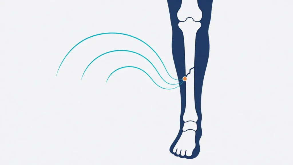
PEMF Osteoporosis: What researchers have studied
Osteoporosis is a condition of reduced bone strength and higher fracture susceptibility. Bone mineral density, or BMD, is one measured part of bone health. A BMD result is not the same as a fracture outcome. The studies below did not establish that an intervention prevents fractures unless fractures were measured, and the studies summarized here did not report fracture-risk outcomes.
The human literature is concentrated in postmenopausal women. Researchers have measured BMD at different skeletal sites, including the lumbar spine, hip, forearm, and femur. They have also measured bone-turnover markers, balance, muscle tests, and pain. These are distinct outcomes. A short-term change in a marker does not demonstrate a lasting BMD change, and a site-specific BMD finding does not automatically describe the rest of the skeleton.
Study designs also differ. Some compare PEMF with sham exposure, some compare it with medication or exercise, and one early study exposed only one forearm. The record includes animal models of estrogen-related bone loss. Those animal studies can help frame biological questions, but they cannot determine what happens in people. The useful question is therefore narrow: what did each study measure, in whom, for how long, and with what comparison?
The studies
Lang et al. 2022 systematic review and meta-analysis
Lang and colleagues reviewed 19 studies involving 1,303 participants with postmenopausal osteoporosis. Their meta-analysis reported pooled BMD differences for some comparisons and sites. In the comparison of PEMF with placebo, the authors reported no statistically significant difference in lumbar-spine BMD. Several bone-turnover markers also showed no statistically significant difference in that comparison.
This review is the broadest source in this page, but its pooled findings combine differing devices, comparators, outcomes, and study designs. Its abstract lists BMD, markers, pain, and adverse events, not fracture outcomes. The pooled BMD results therefore do not answer whether fractures occurred or whether any particular device has the same results.
Lang S, Ma J, Gong S, Wang Y, Dong B, Ma X. “Pulse Electromagnetic Field for Treating Postmenopausal Osteoporosis: A Systematic Review and Meta-Analysis of Randomized Controlled Trials.” Bioelectromagnetics. 2022;43(6):381-393. PMID: 35864717. DOI: 10.1002/bem.22419.
Tabrah et al. 1990
Tabrah and colleagues enrolled 20 women described as osteoporosis-prone. The researchers exposed each participant’s nondominant forearm and used single-photon densitometry to measure the radius before, during, and after the exposure period. They reported a statistically significant increase in bone density in the immediate field area during exposure. That measurement decreased during the following 36 weeks. The untreated arm showed a weaker similar response.
This was a small, localized forearm study rather than a randomized sham-controlled trial. The contralateral-arm pattern also makes the comparison less straightforward. It did not measure fractures, and its temporary, site-specific BMD finding does not establish a whole-skeleton or fracture outcome.
Tabrah F, Hoffmeier M, Gilbert F Jr, Batkin S, Bassett CA. “Bone Density Changes in Osteoporosis-Prone Women Exposed to Pulsed Electromagnetic Fields (PEMFs).” Journal of Bone and Mineral Research. 1990;5(5):437-442. PMID: 2195843. DOI: 10.1002/jbmr.5650050504.
Giordano et al. 2001
This single-blind randomized pilot study assigned 40 outpatients with postmenopausal osteoporosis to PEMF or placebo exposure. BMD was measured at baseline and after three months. The authors reported no statistically significant increase in BMD in either group. In the PEMF group, two serum bone-turnover markers increased during the treatment period and returned to baseline one month after treatment stopped.
The null BMD result is important because biochemical markers and BMD are not interchangeable. The sample was small, the observation period was short, and the report did not measure fractures. It cannot determine whether marker changes corresponded to later BMD or fracture outcomes.
Giordano N, Battisti E, Geraci S, Fortunato M, Santacroce C, Rigato M, Gennari L, Gennari C. “Effect of Electromagnetic Fields on Bone Mineral Density and Biochemical Markers of Bone Turnover in Osteoporosis: A Single-Blind, Randomized Pilot Study.” Current Therapeutic Research. 2001;62(3):187-193. DOI: 10.1016/S0011-393X(01)80030-8.
Liu et al. 2013
Liu and colleagues randomly assigned 44 women with postmenopausal osteoporosis to a course of PEMF or alendronate in an active-controlled trial. They measured lumbar-spine and proximal-femur BMD, lower-extremity manual muscle-test scores, and balance at baseline and through 24 weeks. The authors reported no statistically significant treatment difference between groups for either BMD measure, muscle-test score, or balance score.
An active comparison can show whether groups differed under that study’s conditions. It does not show equivalence to medication, and it does not substitute for a sham-controlled design. The trial was small, had a 24-week horizon, and did not report fractures. Its BMD findings are not evidence about fracture risk.
Liu H-F, Yang L, He H-C, Zhou J, Liu Y, Wang C-Y, Wu Y-C, He C-Q. “Pulsed Electromagnetic Fields on Postmenopausal Osteoporosis in Southwest China: A Randomized, Active-Controlled Clinical Trial.” Bioelectromagnetics. 2013;34(4):323-332. PMID: 23362148. DOI: 10.1002/bem.21770.
Elsisi, Mousa, and ELdesoky 2015
This randomized comparison enrolled 30 women aged 60 to 70 years. Fifteen received low-frequency, low-intensity pulsed magnetic-field sessions and 15 completed circuit weight training. BMD and bone mineral content at lumbar-spine and hip-related sites were measured before and after 12 weeks. The authors reported larger post-treatment BMD changes in the magnetic-field group than in the exercise group at the reported sites.
Both groups received an active intervention, so the trial had no sham condition. The groups and study personnel could not be masked to exercise, the sample was small, and follow-up ended at 12 weeks. It did not measure fractures. Those features limit what the site-specific BMD differences can show.
Elsisi HF, Mousa GSM, ELdesoky MTM. “Electromagnetic Field Versus Circuit Weight Training on Bone Mineral Density in Elderly Women.” Clinical Interventions in Aging. 2015;10:539-548. DOI: 10.2147/CIA.S78485.
Mishima 1988
This is an animal study. Mishima used mature female rats with ovariectomy and sciatic neurectomy to create an osteoporosis model. After long-term systemic PEMF exposure, the author reported no effect on physiologically aged bone or on lost cortical bone in osteoporotic hindlegs. The report did describe greater cancellous-bone volume and bone-formation activity in osteoporotic hindlegs.
The mixed findings matter. This was not a human trial, and its combined surgical model, cage-based exposure, tissue measures, and six-month observation do not match clinical osteoporosis care or consumer-device use. It did not measure human BMD or human fractures.
Mishima S. “The Effect of Long-Term Pulsing Electromagnetic Field Stimulation on Experimental Osteoporosis of Rats.” Journal of UOEH. 1988;10(1):31-45. PMID: 3285429. DOI: 10.7888/juoeh.10.31.
Jing et al. 2013
This is an animal study. Jing and colleagues assigned 30 rats to sham surgery, ovariectomy, or ovariectomy plus PEMF exposure. After 10 weeks, the ovariectomy-plus-PEMF group had higher femoral micro-computed-tomography BMD and several microarchitecture measures than the ovariectomy group. The researchers also observed no obvious effect on RANKL or RANK gene expression.
These findings came from a controlled rat model with laboratory exposures and animal tissue measurements. They do not show a human BMD effect, a fracture outcome, or equivalence between the study equipment and another applicator format.
Jing D, Li F, Jiang M, Cai J, Wu Y, Xie K, Wu X, Tang C, Liu J, Guo W, Shen G, Luo E. “Pulsed Electromagnetic Fields Improve Bone Microstructure and Strength in Ovariectomized Rats Through a Wnt/Lrp5/?-Catenin Signaling-Associated Mechanism.” PLoS One. 2013;8(11):e79377. PMID: 24244491. DOI: 10.1371/journal.pone.0079377.
How strong is this evidence?
The evidence is limited and mixed for BMD in people with postmenopausal osteoporosis. The 2022 systematic review is useful because it gathered a larger body of trials. Yet its pooled comparisons span different devices, field settings, skeletal sites, follow-up periods, co-interventions, and comparators. Its own reported null lumbar-spine result for PEMF versus placebo shows why a single favorable pooled statement cannot represent every outcome or setting.
Individual human studies are small. Tabrah et al. involved 20 participants and a localized forearm measurement. Giordano et al. involved 40 participants and found no statistically significant BMD increase in either group. Liu et al. involved 44 participants and found no statistically significant between-group BMD difference through 24 weeks. Elsisi et al. compared two active interventions in 30 participants without a sham group. These designs leave uncertainty from small samples, short follow-up, blinding limitations, and differences in baseline care and measurement sites.
There is also a direct outcome gap. BMD is not fracture risk, even though the two are related in osteoporosis assessment. The studies summarized here did not report fracture outcomes. The animal studies add controlled laboratory observations and include null findings, but animal models cannot resolve human effectiveness, safety, or long-term outcomes. This record supports careful reporting of named measurements. It does not support conclusions about fracture prevention or replacement of prescribed osteoporosis care.
What this means if you are considering a PEMF device
Keep the research question separate from a product decision. Ask whether a study used a localized forearm, a trunk or hip-area application, or a laboratory animal enclosure. A portable or localized applicator may resemble the anatomical placement in a localized study more closely than a full-body mat. That is a format comparison only. It is not evidence of equivalence, a recommendation, or a reason to expect the same result from another device.
Compare written specifications, the intended general-wellness positioning, return terms, and documentation before buying. For general equipment context, compare the written descriptions of portable applicators and full-body mats. Osteoporosis management and prescribed treatment decisions remain matters for the clinician who knows your health history.
Questions to ask before buying
- Does the seller provide the waveform, frequency range, field-strength information, and applicator dimensions in writing?
- Does the research summary identify the study population, skeletal site, comparison group, and whether fractures were measured?
- Is the applicator format described clearly enough to compare its placement with the placement used in a cited study?
- What are the written return window, warranty terms, repair process, and any restocking fees?
- Are the manual, contraindication information, and product limitations available before purchase?
This page summarizes published research for reference. It is not medical advice, and nothing here is a claim that any PEMF device treats, cures, or prevents any condition. PEMF is not appropriate for everyone — review PEMF contraindications and talk with your own physician before beginning any new therapy, particularly if you have an implanted electronic device, are pregnant, or are being treated for a serious medical condition.