
This page summarizes published research on electromagnetic-field exposures and wound-related outcomes. It is part of ElectroMeds’ PEMF education library and does not state what any device will do for a wound or a person with a wound-related condition.
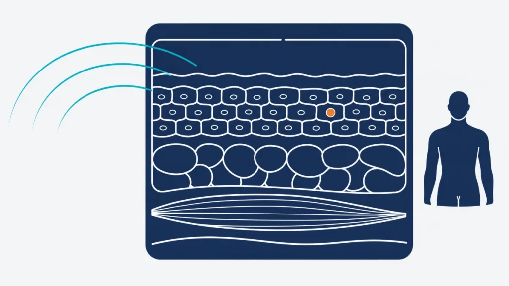
PEMF Wound Healing: What researchers have studied
Wound-healing research is not one uniform body of work. The human studies summarized here involve chronic venous leg ulcers, pressure ulcers in people with spinal cord injuries or neurological disorders, and chronic diabetic foot ulcers. Researchers have measured different outcomes, including change in wound area, time to complete closure, wound-depth measures, and investigator assessments. Some experiments used pulsed electromagnetic energy or radio-frequency pulsed energy rather than the low-frequency PEMF formats commonly discussed by consumers. Those differences matter when comparing studies.
The literature also includes an animal experiment on standardized rat skin wounds. That type of work can test a controlled model, but it cannot establish what happens in people. Across the human research, electromagnetic exposure was studied alongside wound dressings or conventional care, not as a substitute for clinical evaluation. The reviews and trials below include positive findings and a later randomized trial that found no statistically significant between-group difference.
The original page also listed historical records that do not have a traceable PMID or DOI, refer to static magnets, or address another condition. They are preserved and flagged in the hidden preservation audit rather than used as visible evidence.
The studies
Chronic-wound systematic review
McGaughey and colleagues systematically reviewed controlled clinical studies of pulsed electromagnetic energy in chronic ulcers. They included 11 studies with 360 participants across venous, pressure, and plantar ulcers. The review reported that most sham-controlled venous-ulcer studies found a statistically significant difference favoring active exposure, while evidence for pressure and plantar ulcers was more limited. The review did not pool a single effect estimate because the field strengths, schedules, devices, wound types, and outcome measures varied substantially. Its findings therefore describe a mixed collection of interventions, not one defined PEMF device or format.
McGaughey H, Dhamija S, Oliver L, Porter-Armstrong A, McDonough S. “Pulsed electromagnetic energy in management of chronic wounds: a systematic review.” Physical Therapy Reviews, 14(2), 132-146, 2009. DOI: 10.1179/174328809X435231.
Cochrane review of pressure ulcers
Aziz and Bell-Syer reviewed randomized trials of electromagnetic therapy for pressure ulcers. The review included two trials totaling 60 participants, both judged at unclear risk of bias. Neither trial found a statistically significant difference in complete healing compared with the control group. One study reported a statistically significant difference in percentage reduction in wound area, but the reviewers cautioned that this was a small study and that area reduction is less clinically meaningful than complete healing. This is an important counterweight to more favorable reports. The review addressed electromagnetic therapy broadly, including electrode-based approaches, so it is not a direct evaluation of every PEMF format.
Aziz Z, Bell-Syer SEM. “Electromagnetic therapy for treating pressure ulcers.” Cochrane Database of Systematic Reviews, 2015(9), CD002930, 2015. PMID: 26334539. DOI: 10.1002/14651858.CD002930.pub6.
Recalcitrant venous-ulcer trial
Stiller and colleagues conducted a prospective, multicenter, randomized, double-blind, placebo-controlled trial in people with recalcitrant, predominantly venous leg ulcers. At eight weeks, the active-exposure group had a 47.7% decrease in wound surface area, while the placebo group had a 42.3% increase; the article reported a statistically significant between-group difference. Wound-depth and pain measures also favored the active group in this trial. The exposure was used with a wound dressing, so the result does not isolate a consumer device from clinical wound care. The abstract does not establish whether a different device design, output, wound cause, or care setting would yield the same result.
Stiller MJ, Pak GH, Shupack JL, Thaler S, Kenny C, Jondreau L. “A portable pulsed electromagnetic field (PEMF) device to enhance healing of recalcitrant venous ulcers: a double-blind, placebo-controlled clinical trial.” British Journal of Dermatology, 127(2), 147-154, 1992. PMID: 1390143. DOI: 10.1111/j.1365-2133.1992.tb08047.x.
Primary-care venous-leg-ulcer pilot trial
Kenkre and colleagues enrolled 19 people with venous leg ulcers in a prospective randomized, double-blind controlled pilot trial. At day 50, the group assigned to one electromagnetic setting had significantly greater ulcer healing and lower pain scores than the placebo group or a second active-setting group. The study also reported that all participants improved in mobility by the end of the trial. The small sample is an immediate limitation, and the comparison of two active settings complicates a simple active-versus-sham interpretation. As with the Stiller trial, the exposure was an adjunct to conventional dressings and involved a particular clinical population.
Kenkre JE, Hobbs FD, Carter YH, Holder RL, Holmes EP. “A randomized controlled trial of electromagnetic therapy in the primary care management of venous leg ulceration.” Family Practice, 13(3), 236-241, 1996. PMID: 8671131. DOI: 10.1093/fampra/13.3.236.
Pressure-ulcer trial in spinal cord injury
Salzberg and colleagues ran a randomized, double-blind study in 30 male patients with spinal cord injuries and stage II or III pressure ulcers. Among those with stage II ulcers, the active-exposure group had a greater percentage of the ulcer healed at one week, and active exposure was associated with a shorter median time to complete healing after adjustment for baseline ulcer status. Stage III ulcers also healed faster in the active group, according to the authors, but the stage III sample was limited. The all-male spinal-cord-injury population and pressure-ulcer setting constrain generalizability to other wounds. The report’s result cannot determine how a different device, exposure method, or wound-care environment would compare.
Salzberg CA, Cooper-Vastola SA, Perez F, Viehbeck MG, Byrne DW. “The effects of non-thermal pulsed electromagnetic energy on wound healing of pressure ulcers in spinal cord-injured patients: a randomized, double-blind study.” Ostomy Wound Management, 41(3), 42-48, 1995. PMID: 7546114.
Pressure-ulcer trial with a null between-group result
Gupta and colleagues conducted a randomized, double-blind, sham-controlled trial involving 12 people with neurological disorders and 24 pressure ulcers. Both the active and sham groups had statistically significant improvement from baseline on the study’s wound-assessment score. When the groups were compared with each other, however, the differences were not statistically significant for either the Bates-Jensen wound assessment score or the pressure-ulcer staging score. This direct null result is especially relevant because it used a sham comparison. The trial was very small, counted ulcers as well as participants, and assessed one particular whole-body exposure arrangement, which limits precision and relevance to other formats.
Gupta A, Taly AB, Srivastava A, Kumar S, Thyloth M. “Efficacy of pulsed electromagnetic field therapy in healing of pressure ulcers: a randomized control trial.” Neurology India, 57(5), 622-626, 2009. PMID: 19934563. DOI: 10.4103/0028-3886.57820.
Experimental cutaneous wounds in rats
Patiño and colleagues performed an animal study in 22 male Wistar rats with standardized circular skin wounds. The researchers compared a sham group, a topical nitrofurazone group, and a pulsed-electromagnetic-field group. Wound area and perimeter were lower in the electromagnetic-field group than in the sham group at days 7, 14, and 21; the difference from the nitrofurazone group was reported at day 21 only. These observations are relevant to a laboratory skin-wound model, not to clinical decision-making for people. Rat skin, induced wounds, comparator care, and experimental conditions differ from the many causes and complexities of human wounds.
Patiño O, Grana D, Bolgiani A, Prezzavento G, Miño J, Merlo A, Benaim F. “Pulsed electromagnetic fields in experimental cutaneous wound healing in rats.” Journal of Burn Care & Rehabilitation, 17(6 Pt 1), 528-531, 1996. PMID: 8951540. DOI: 10.1097/00004630-199611000-00009.
How strong is this evidence?
The evidence is suggestive in some settings but remains uneven. The better-known human trials used randomization and blinding, and several reported favorable wound-area or healing-time results in chronic venous or pressure ulcers. Yet the positive studies were generally small, were conducted decades ago, and used distinct electromagnetic systems. The 2009 systematic review found 11 trials with only 360 participants in total and reported methodological scores ranging from 1 to 9 out of 11. It also found major variation in devices, exposure schedules, ulcer causes, and endpoints. Those differences make it difficult to combine results or infer an effect for a different device.
The mixed record matters. In the Gupta trial, both active and sham groups improved, with no statistically significant difference between them. The Cochrane review found no statistically significant difference in complete pressure-ulcer healing in either of its two included trials and concluded that strong evidence was lacking. Conversely, the Salzberg pressure-ulcer study and the Stiller and Kenkre venous-ulcer trials reported differences favoring their active interventions. This is not a settled, replicated evidence base.
The conditions also are not interchangeable. Venous leg ulcers, pressure ulcers, postsurgical bruising, diabetic foot ulcers, and experimental skin wounds have different causes, risks, clinical priorities, and usual care. Much of the older literature used pulsed electromagnetic energy or pulsed radio-frequency systems, while some legacy sources concern static magnets. A result in one wound type and device configuration does not establish whether it applies to another. Follow-up was often short, and complete healing, recurrence, and patient-important outcomes were not measured consistently.
What this means if you are considering a PEMF device
The research above may help a reader understand what researchers tested, but it does not identify a consumer device for wound care. A wound needs clinical assessment because its cause, depth, circulation, infection risk, pressure exposure, and management needs can differ substantially. Published research does not replace that assessment or prescribed care.
Format relevance is a comparison question, not evidence of equivalence. For a localized area of interest, a localized-applicator format most closely resembles the positioning used in several localized studies, whereas whole-body arrangements resemble a different study context. The output, waveform, field strength, duration, and device construction still may not match, and the studies do not establish whether results transfer between formats. The PEMF science page and what PEMF is provide background for evaluating those descriptions without turning a research finding into a personal outcome claim.
Questions to ask before buying
- Does the seller clearly identify the applicator size, intended positioning options, and included accessories?
- Are the device’s listed waveform, frequency range, field-strength information, and power source documented in writing?
- Does the seller distinguish product specifications from findings in named studies?
- What are the warranty terms, return conditions, and any restocking fees?
- Is the user manual available before purchase, including cleaning, storage, and maintenance instructions?
This page summarizes published research for reference. It is not medical advice, and nothing here is a claim that any PEMF device treats, cures, or prevents any condition. PEMF is not appropriate for everyone — review PEMF contraindications and talk with your own physician before beginning any new therapy, particularly if you have an implanted electronic device, are pregnant, or are being treated for a serious medical condition.